Images that help you show the best aspects of your product
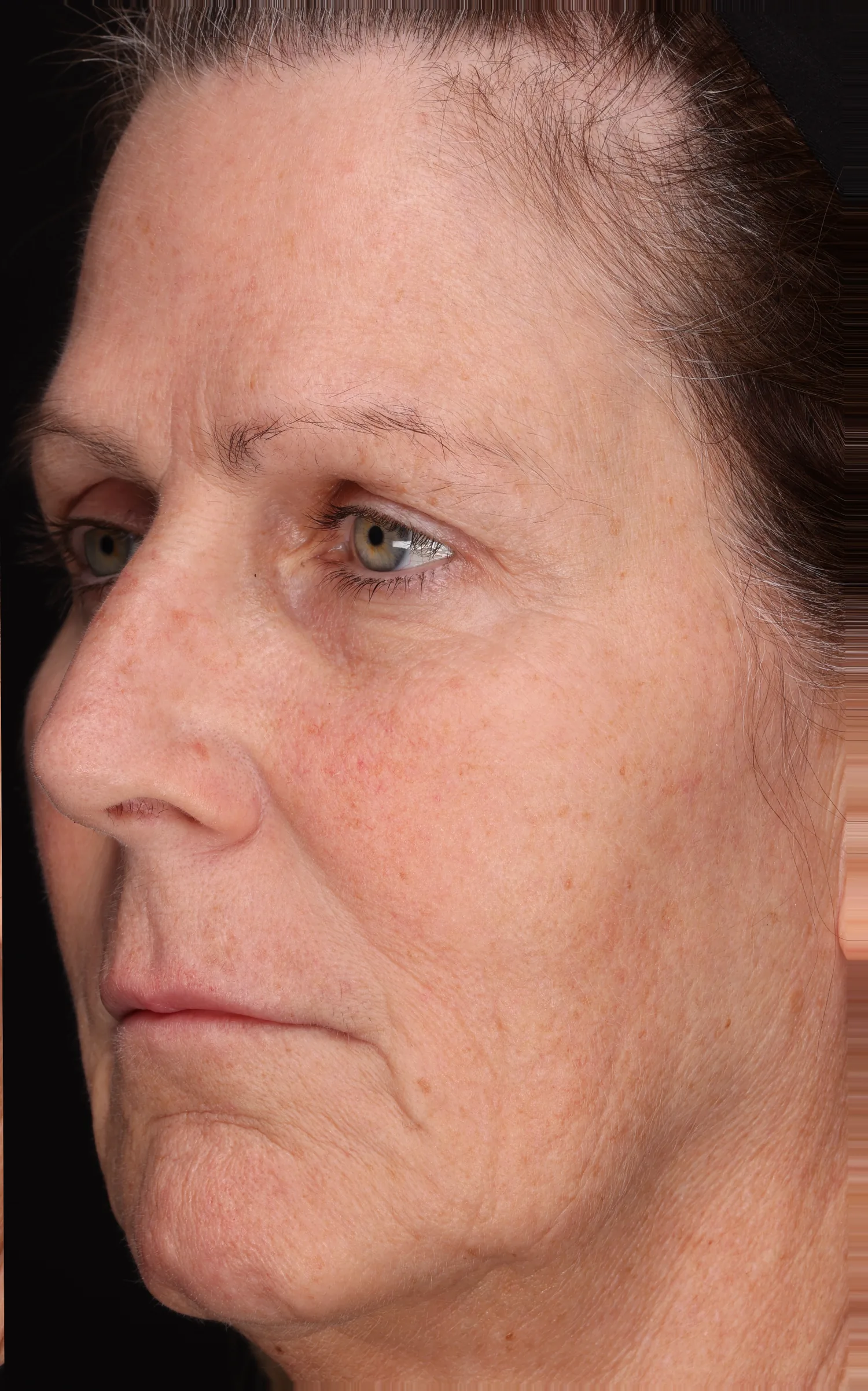
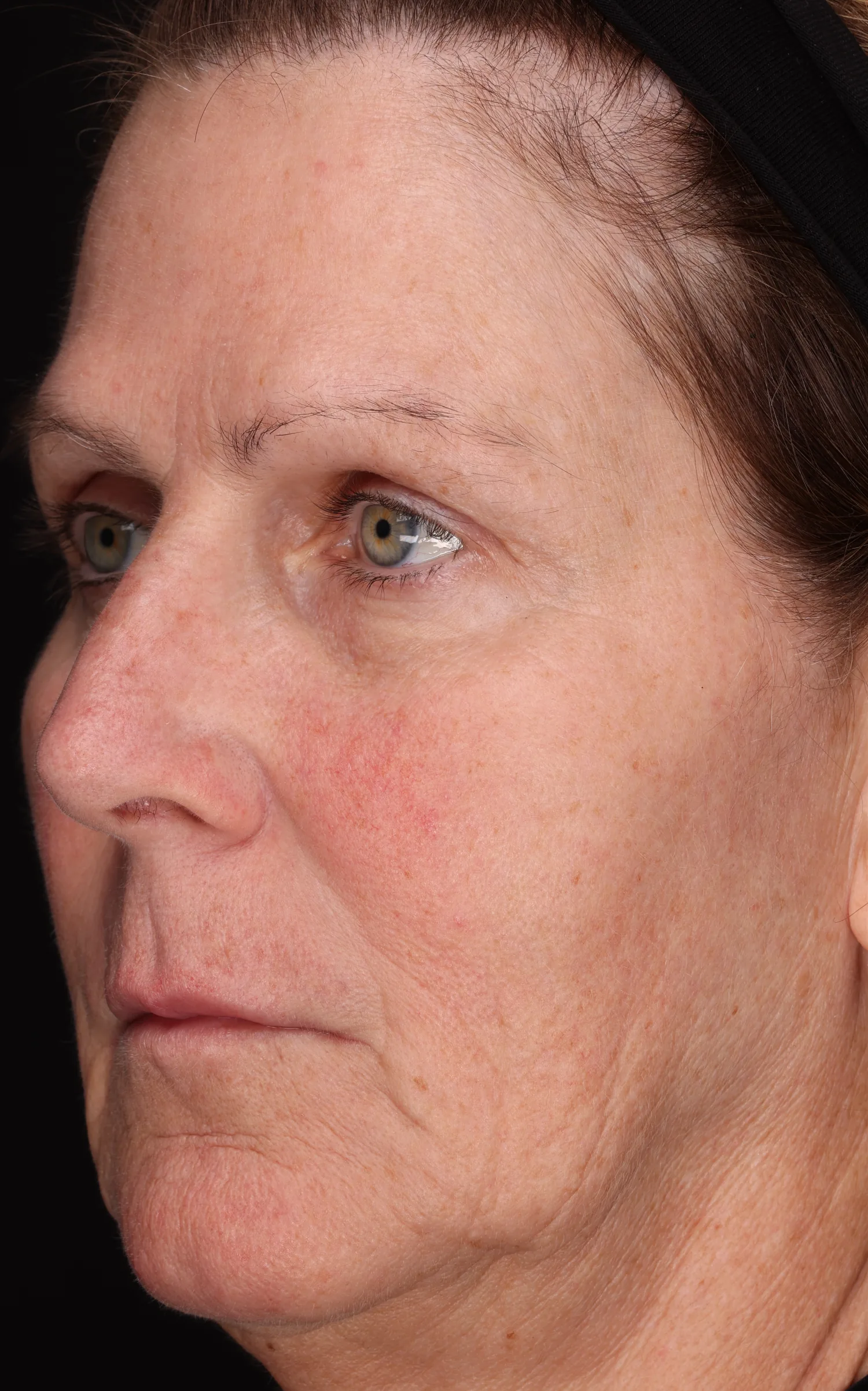
Every study image is captured under controlled light at extreme resolution, so a single frame can fill a tradeshow booth wall or zoom to the edge of a fine line in a digital marketing package. This page shows the same images working on screen, on a website, at a trade show, in print and in multiple styles of image analysis.
Clinical photography gallery
- 25.4 MP: per frame, 4,271 × 5,955 pixels
- True color: no retouching, no smoothing
- Same lighting, same positioning, every visit: the only variables are product efficacy and time
- Native resolution: no upscaling
- Measured in pixels: each result is a defined mathematical count
- Visits lined up: baseline and follow-up matched frame to frame down to the pixel
- Face and body: from crow’s feet to cracked heels
- One image, multiple formats: ready for web, print, TV and tradeshow material
- Analysis you can switch on: overlays show exactly what was measured
- Shot in Teaneck, NJ: in a temperature and humidity controlled studio
From the whole face to the crow’s feet, in one move
Scroll to zoom. Both visits zoom together, so the change stays side by side the whole way in.
A slider your customers can drag themselves
Drop the comparison onto a product page. Shoppers drag to see the change, and one tick lays the image analysis over the photo so they can see what was measured.
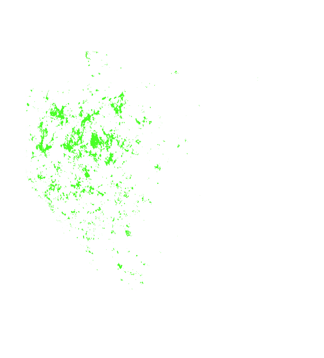
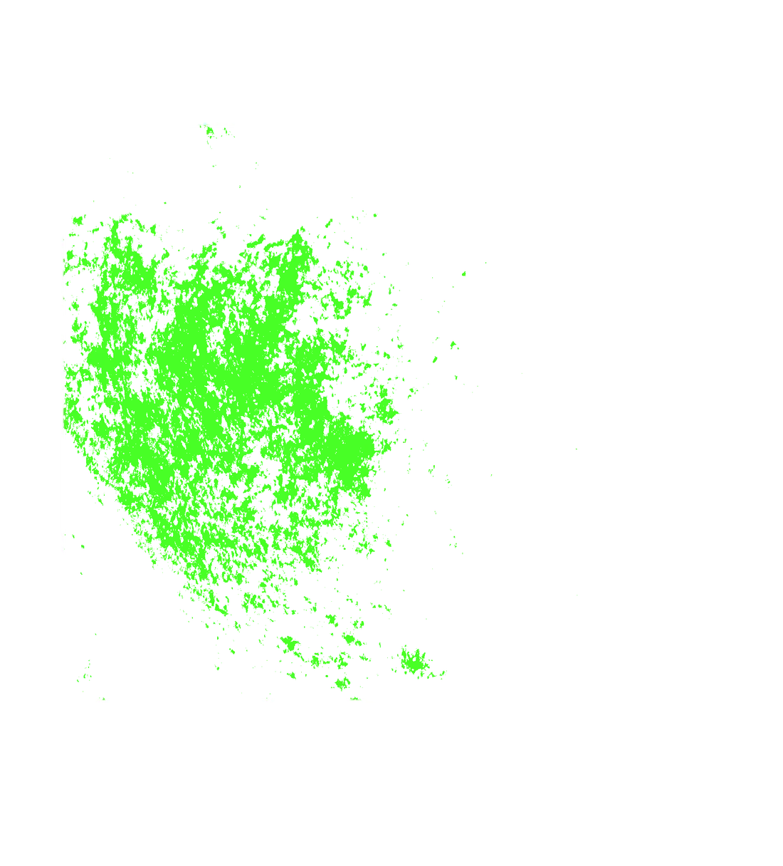
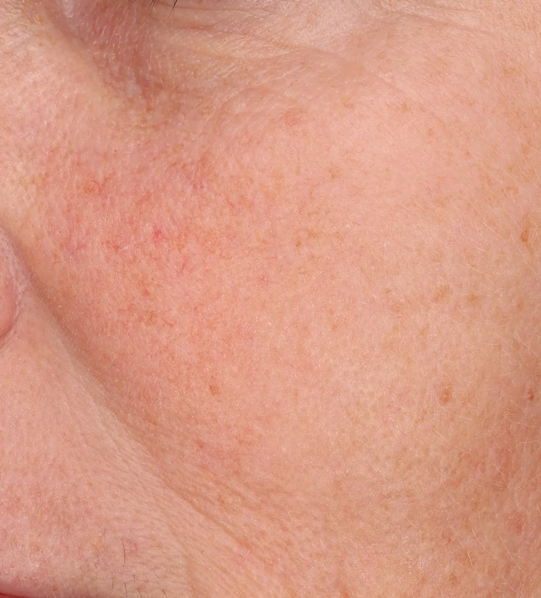
Drag across the photo to compare. Tick the box to show the red pixels that were measured, 79% fewer by week 2.
Side by side full resolution before and afters, with zoomed in sub-regions.
Baseline and week 4 hang side by side. A thin ring marks the patch of cheek on each face, and the magnified circle sits in the banner’s text panel, so the lesions stay visible on the photo and up close.
Matched close-ups Both circles are cropped from their own frames at the same measured location.
Built for print No hover, no motion. Everything a visitor needs is on the banner itself.
Zoom one visit, and the other follows
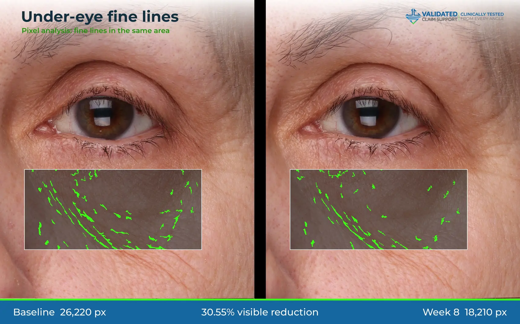
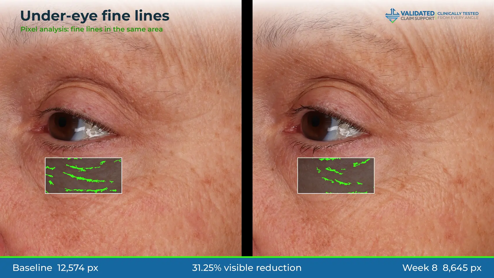
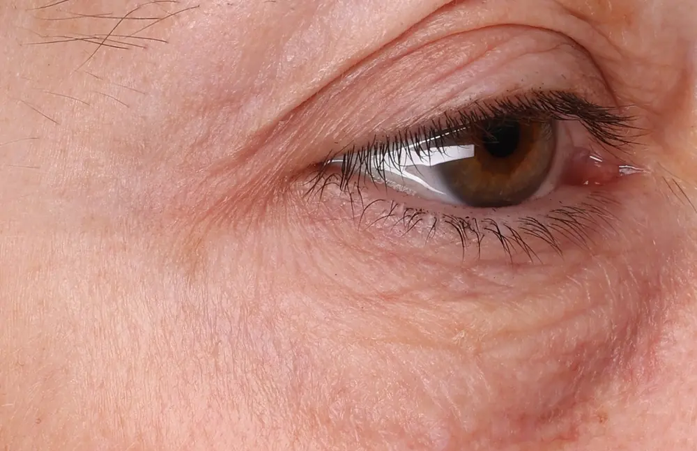
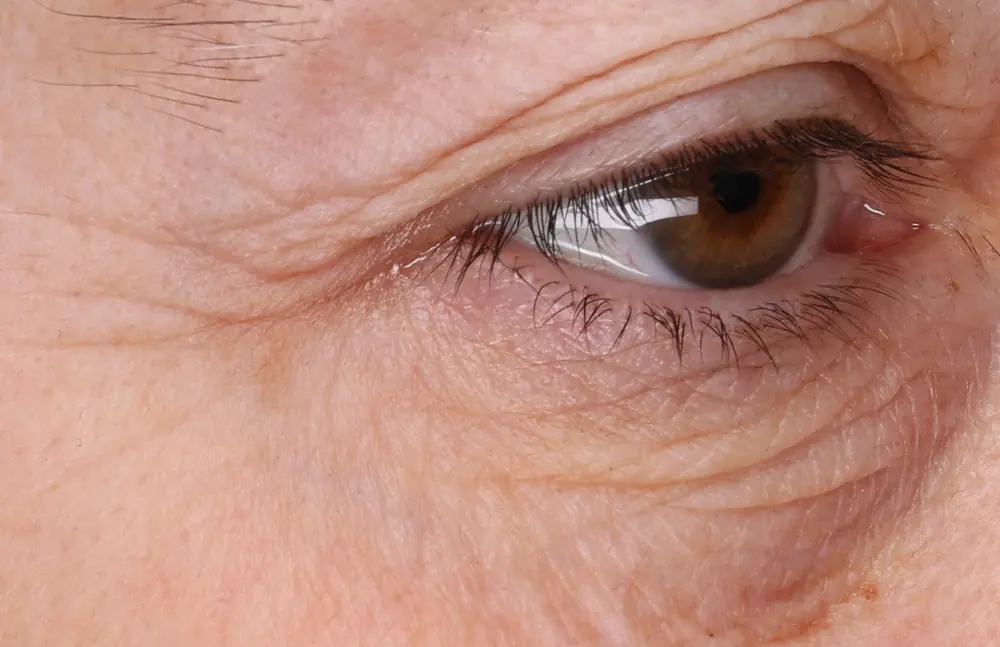
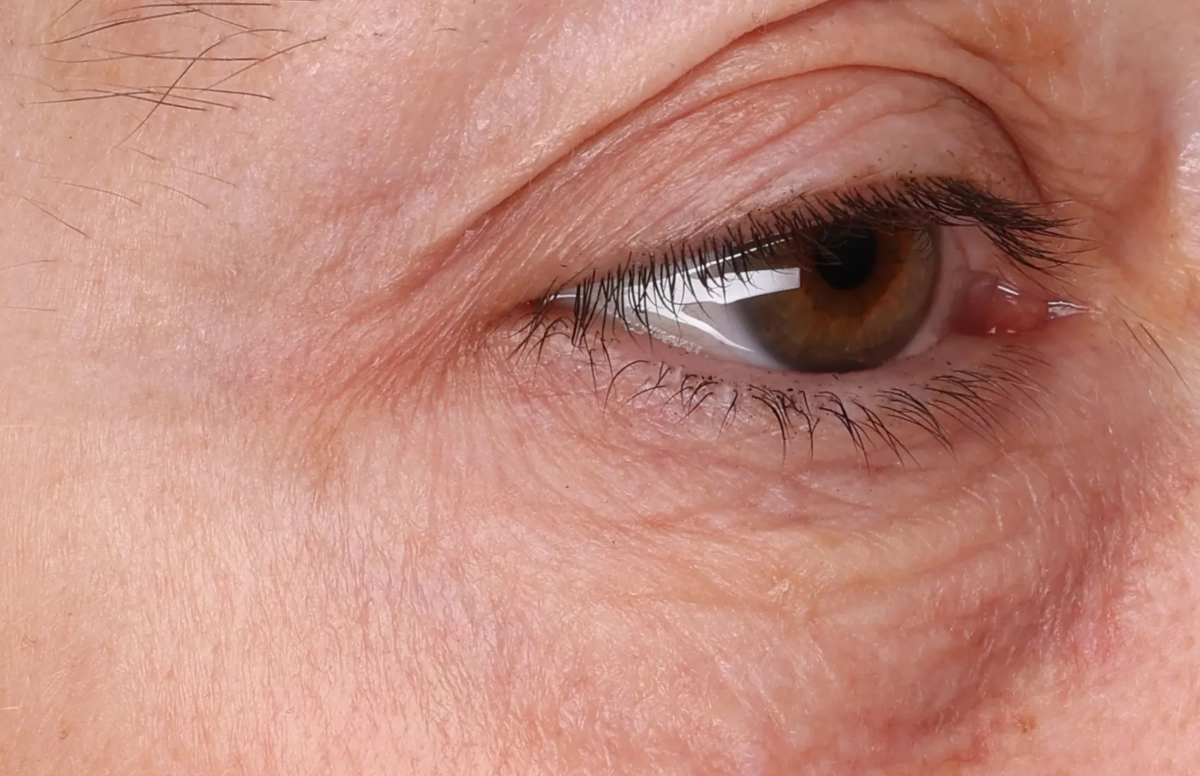
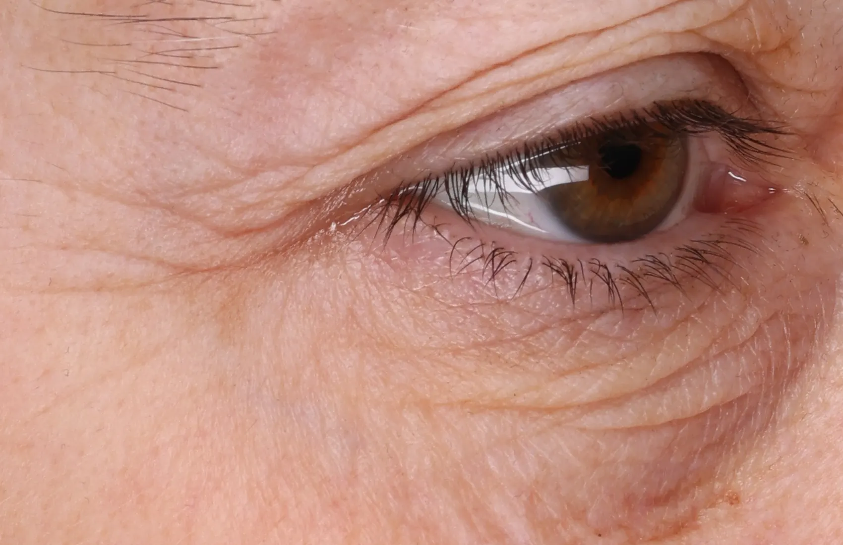
Scroll, pinch or drag either image and both move together. On a computer, rest the cursor on either photo and a zoom circle appears on both, at the same spot. Fine lines around the eye, baseline to week 8.
Before, after and the measurement behind them
Finished case study pages, ready for a brochure or sell sheet. Each one carries the photos, the pixel analysis and the measured change, so a reader can check the claim on the page. Where a case comes in more than one format, both are shown.
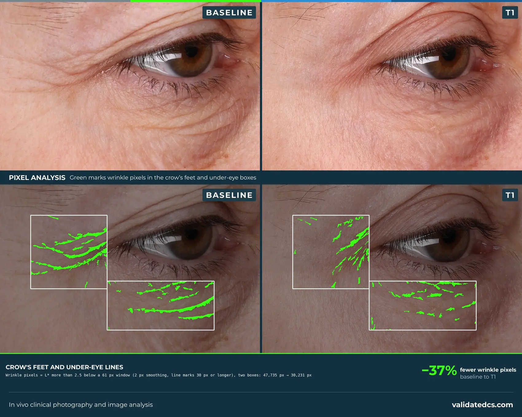
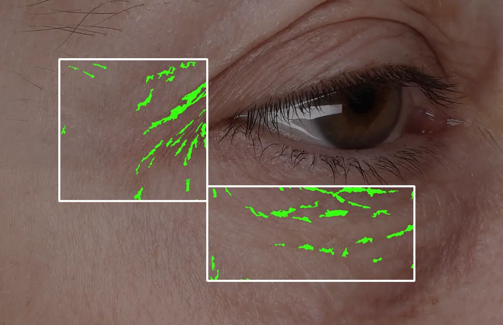
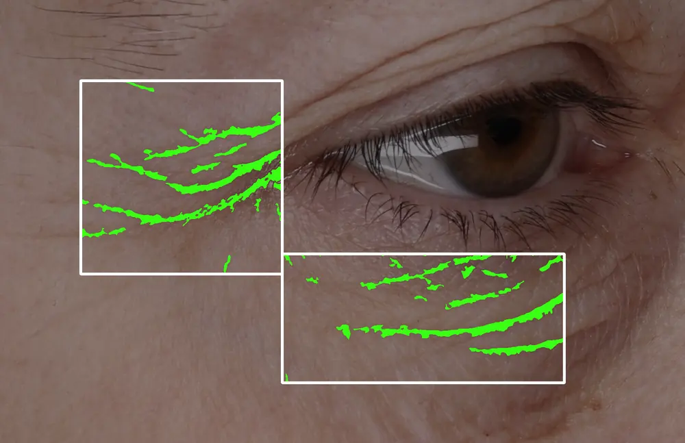
Crow’s feet and under-eye −37% wrinkle pixels, baseline to T1. Case study layout.
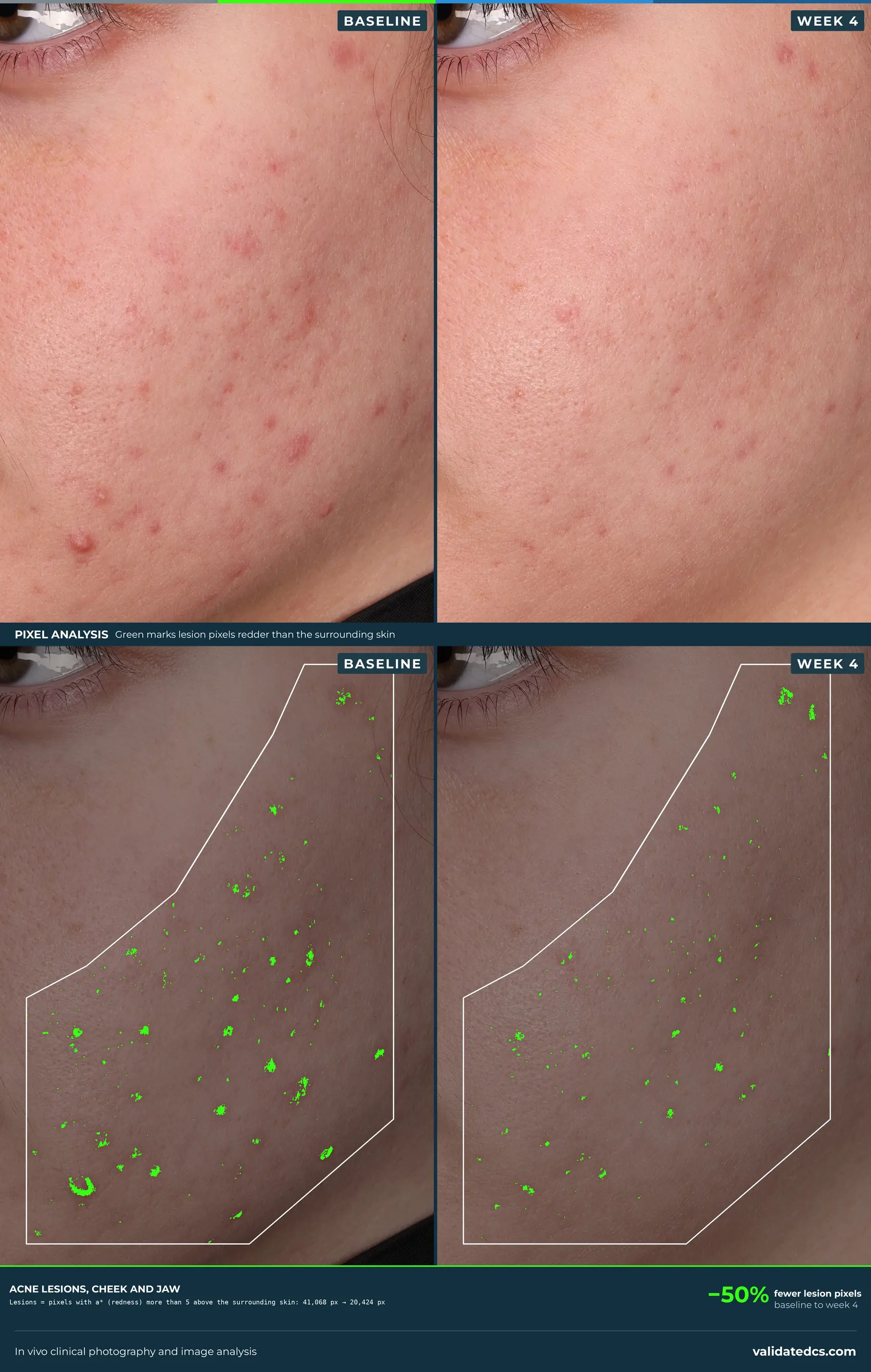
Acne lesions, cheek and jaw −50% lesion pixels, baseline to week 4. Case study layout.
Redness, cheek −79% red pixels, baseline to week 2. Case study layout.
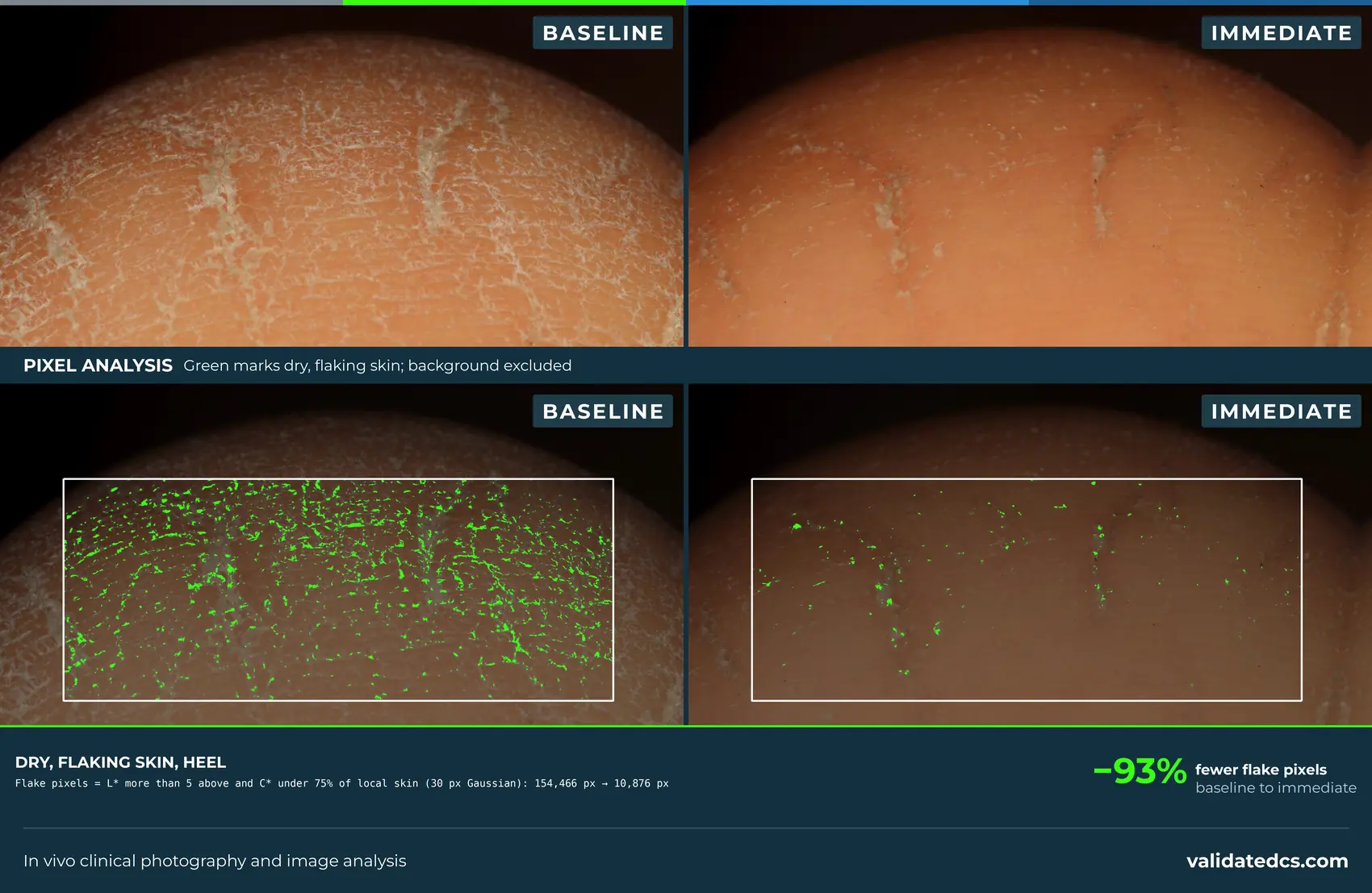
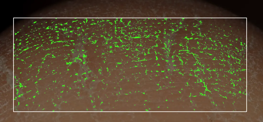
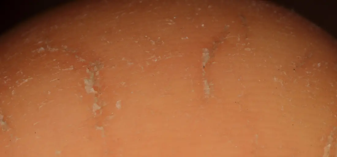
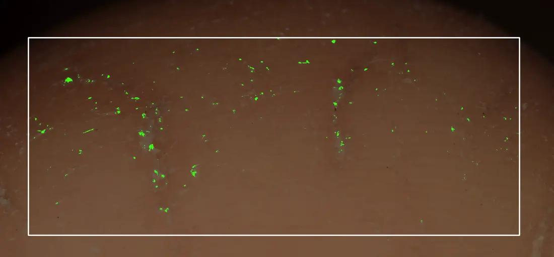
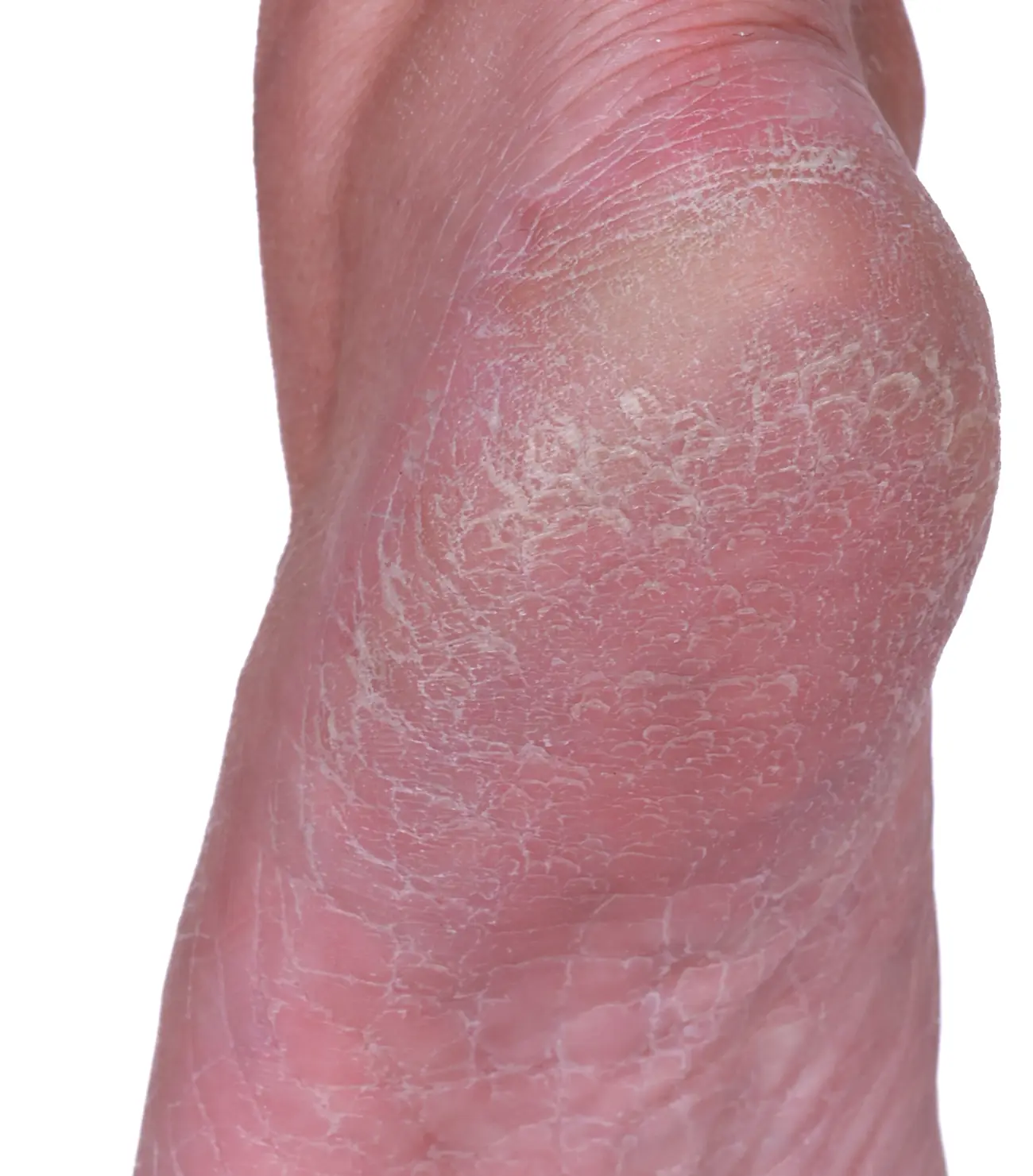
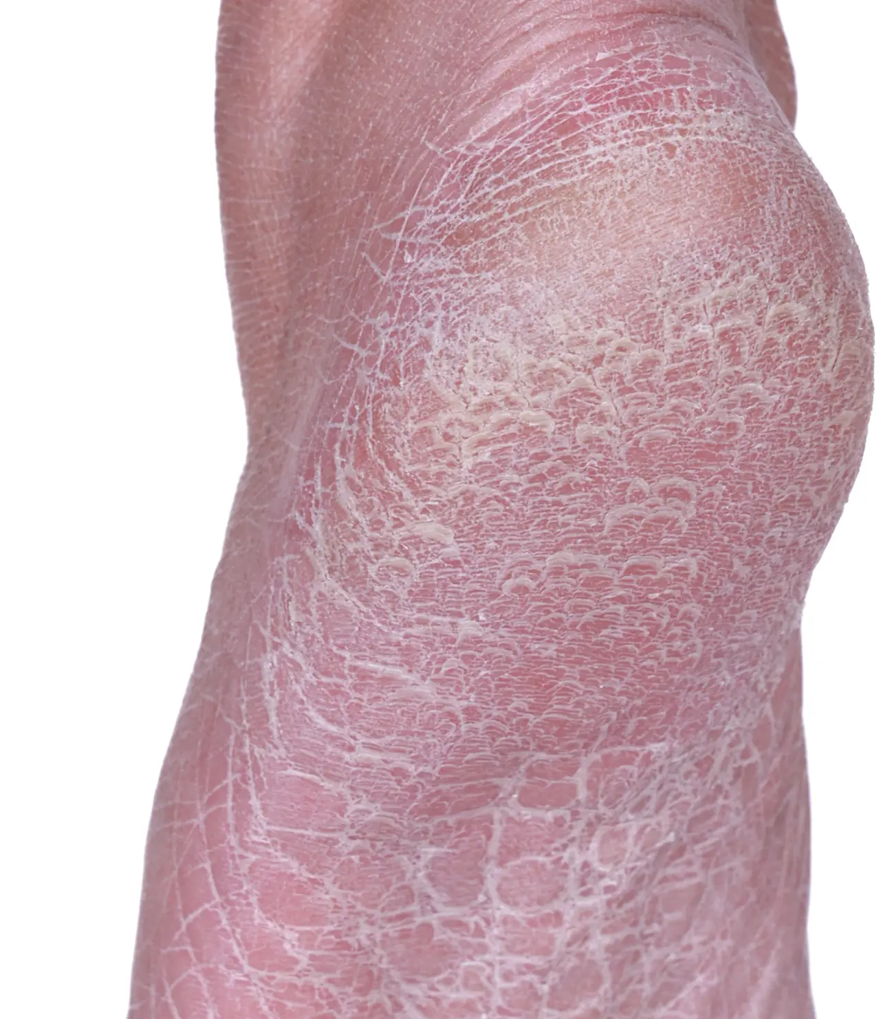
Dry, flaking heel −93% flake pixels, baseline to immediately after. Case study layout.
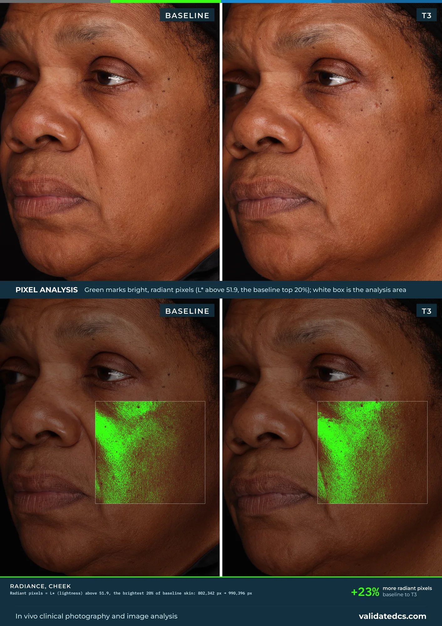
Radiance, cheek +23% more radiant pixels, baseline to T3. Print layout with pixel analysis.
Radiance, cheek +23% more radiant pixels, baseline to T3. 16:9 screen layout.
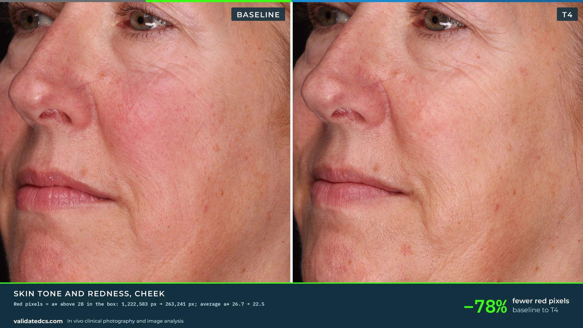
Skin tone and redness, cheek −78% fewer red pixels, baseline to T4. Print layout with pixel analysis.
Skin tone and redness, cheek −78% fewer red pixels, baseline to T4. 16:9 screen layout.
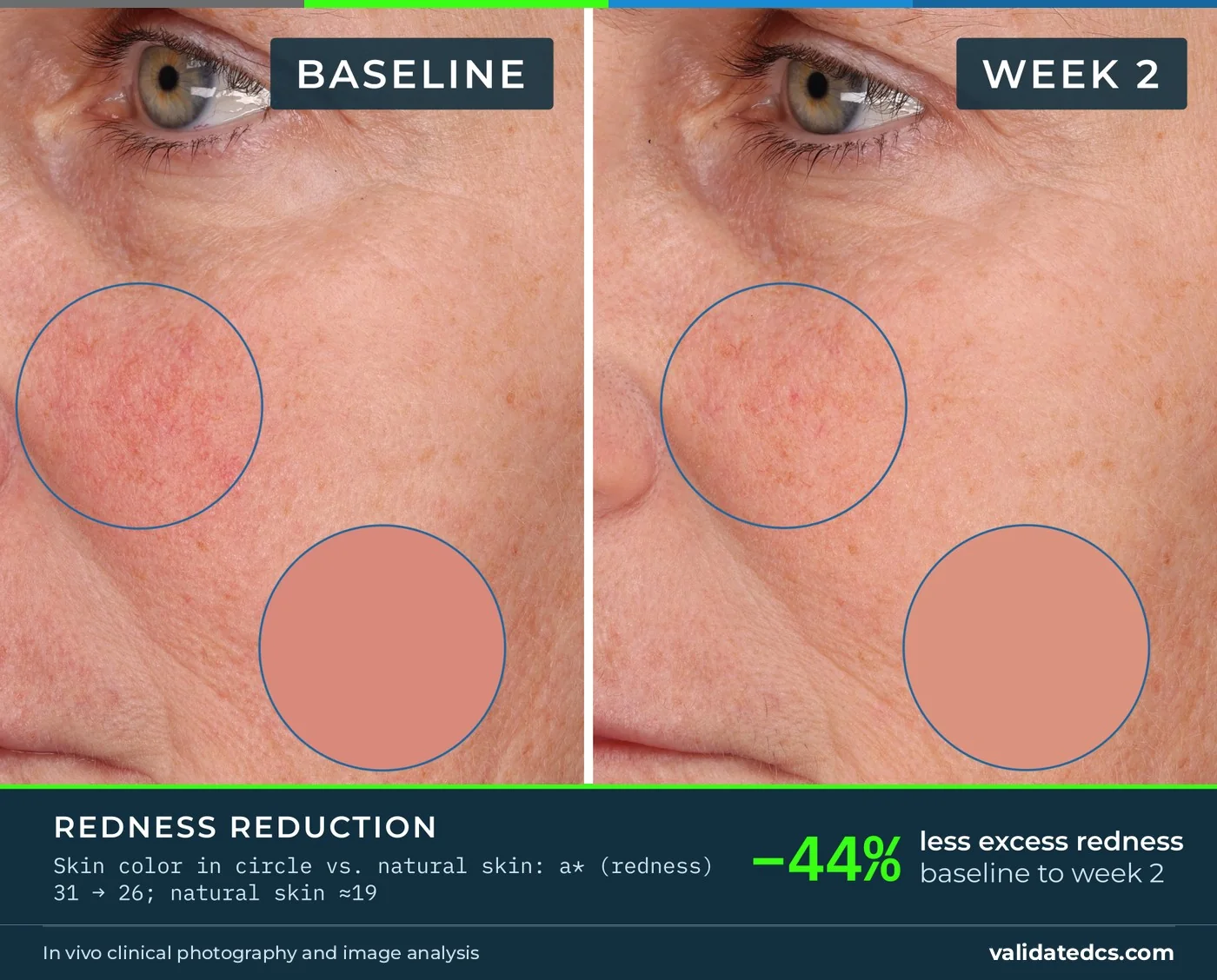
Redness reduction, cheek −44% less excess redness, baseline to week 2. Color swatch layout.
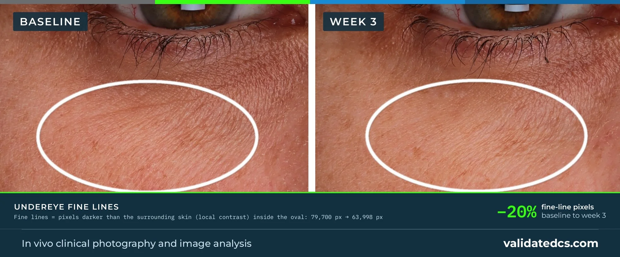
Under-eye fine lines −20% fine-line pixels, baseline to week 3. Print layout with pixel analysis.
Under-eye fine lines −20% fine-line pixels, baseline to week 3. Photos only.
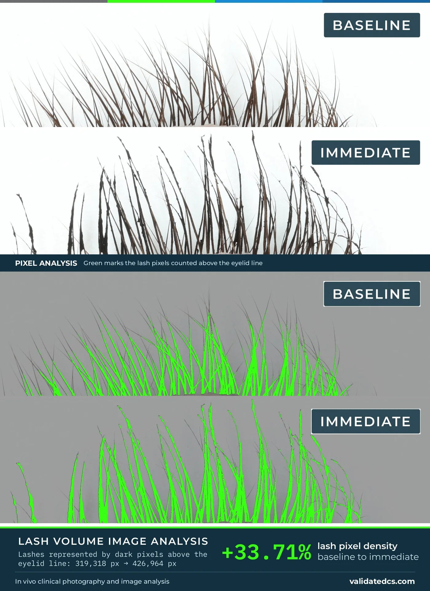
Lash volume +33.71% lash pixel density, baseline to immediate. Print layout with pixel analysis.
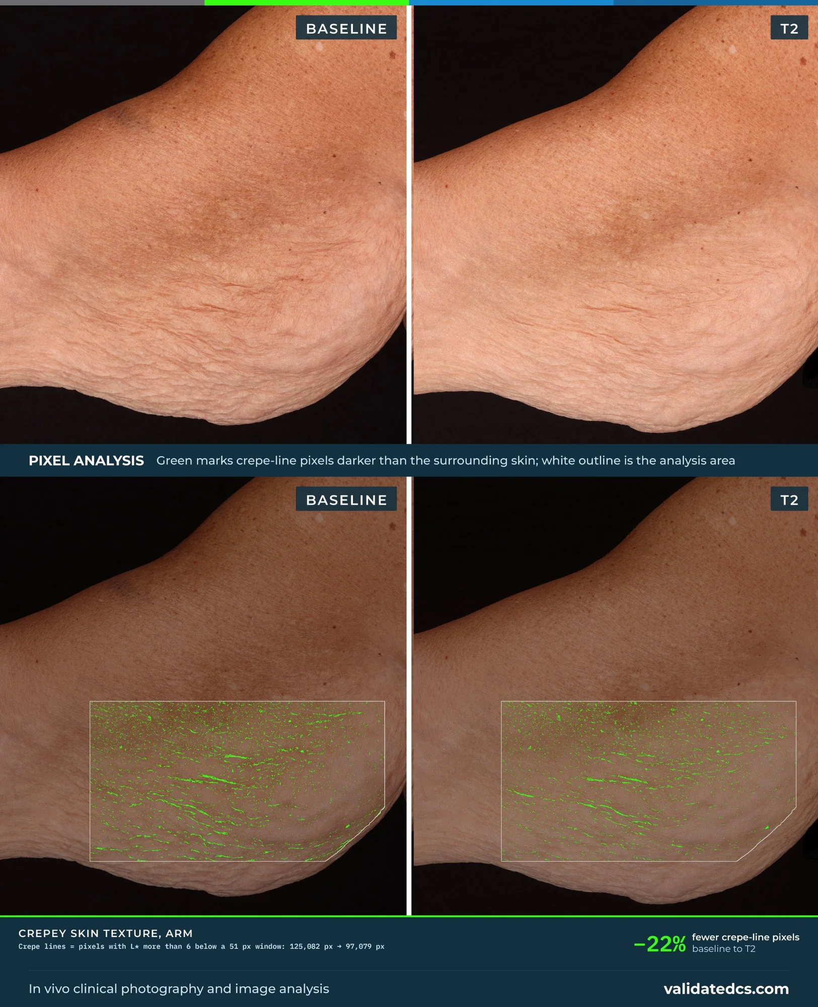
Crepey skin texture, arm −22% fewer crepe-line pixels, baseline to T2. Print layout with pixel analysis.
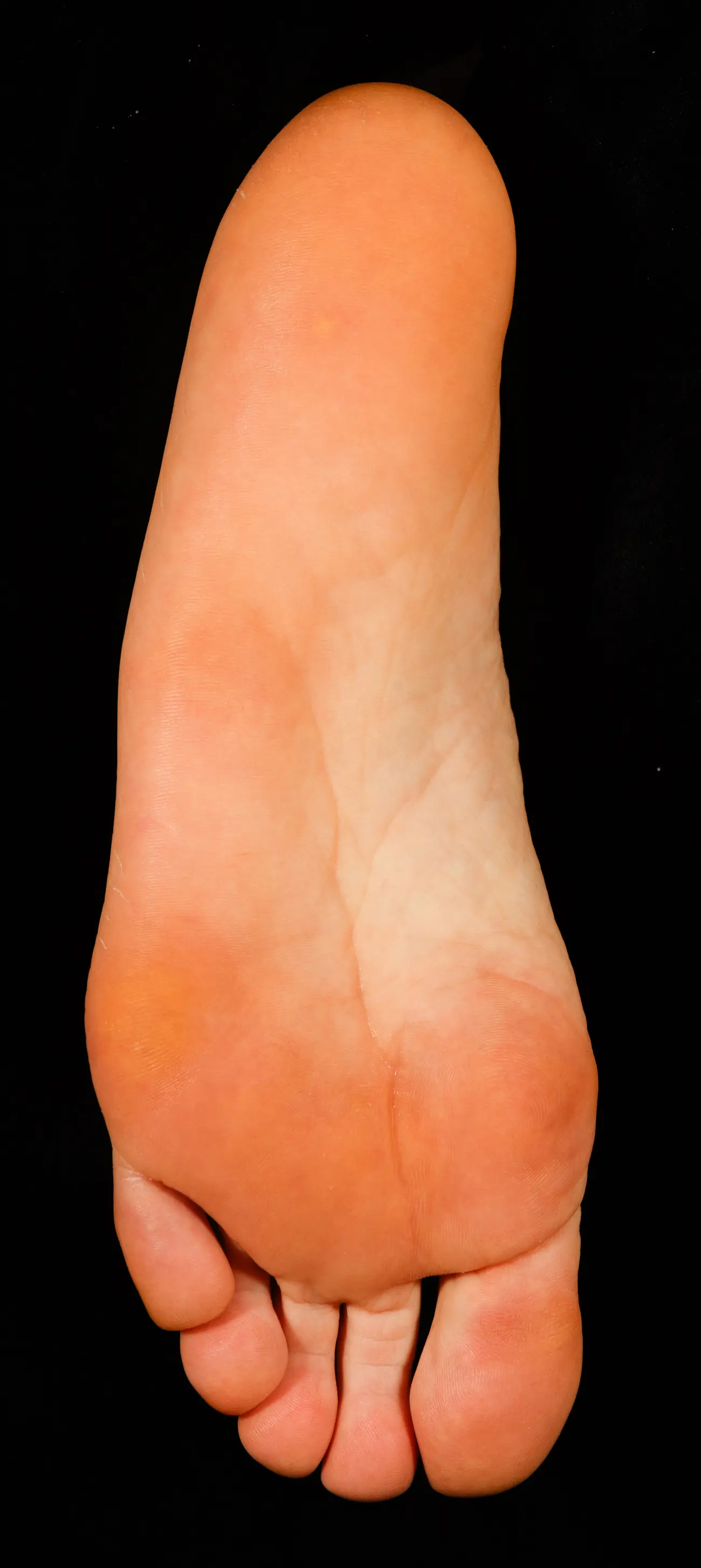
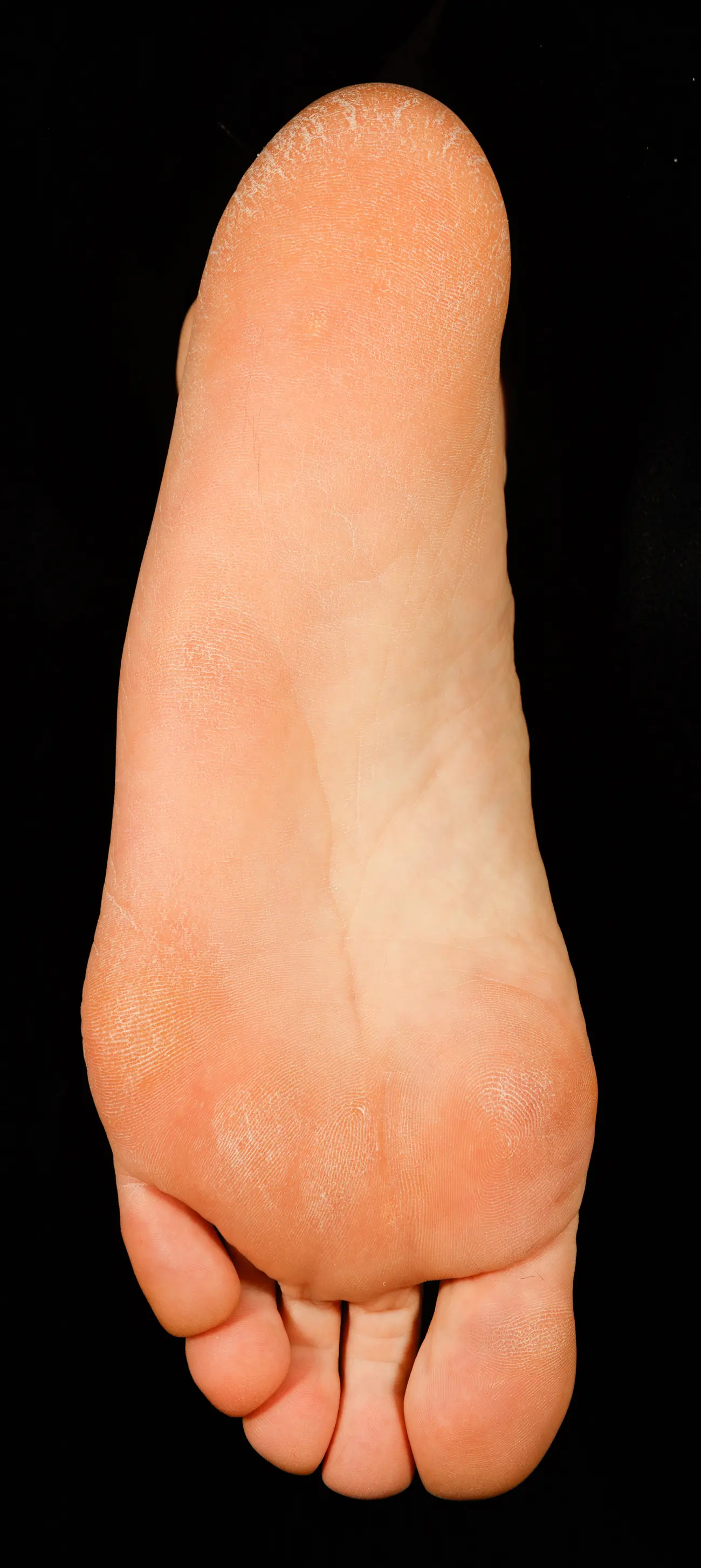
From the whole foot to the cracked heel closeup
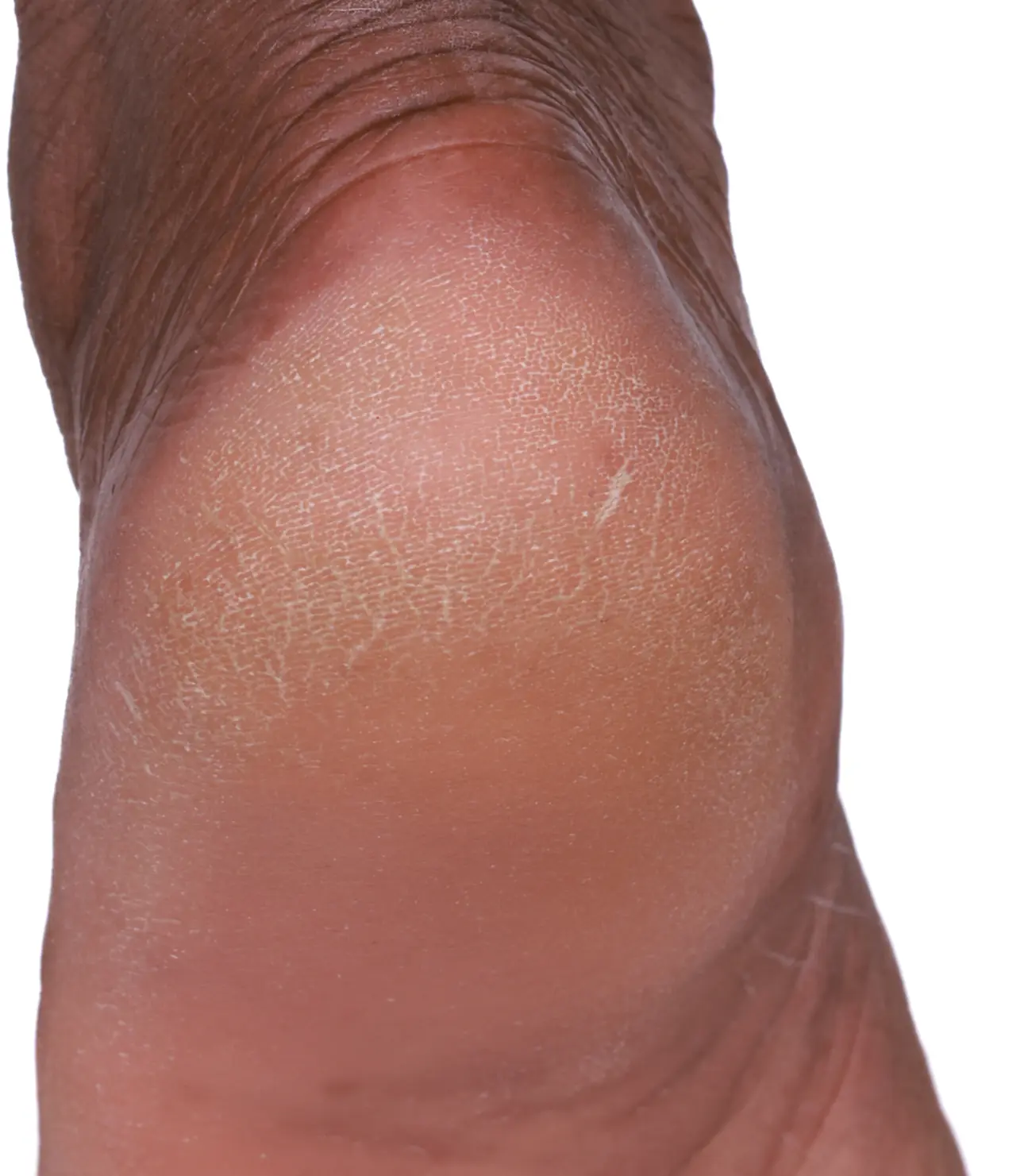
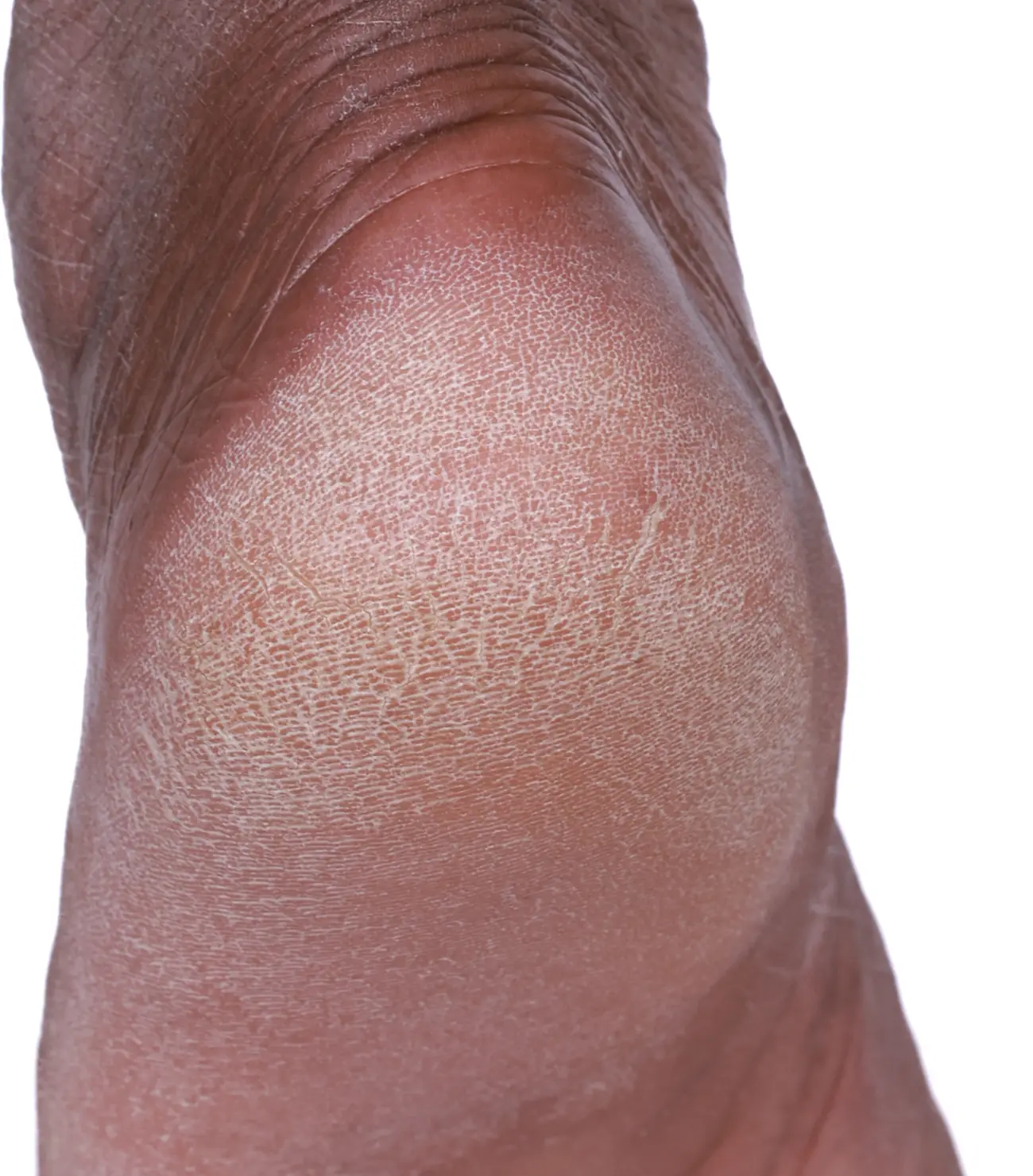
The same move works on the body. Keep scrolling to land on the dry heel, before and after one application.
Drag to compare, or open full screen
Heels, hands and eczema patches at a moderate zoom. Drag across each photo to compare, or open it full screen to zoom further and see the analysis where there is one.
Pick the style that fits the job
Custom outputs ready for multiple print and digital formats.
Open any image full screen
Rest on a tile to see the change. Open it to zoom to full resolution, compare visits, switch to the analysis, and move through the other images from the same case.
Want images like these for your product?
Clinical photography and image analysis can be part of any VCS study, ready for your website, booth, deck and print.
Body care cases
- Cracked heel: Baseline to after, 72.74% fewer flake pixels
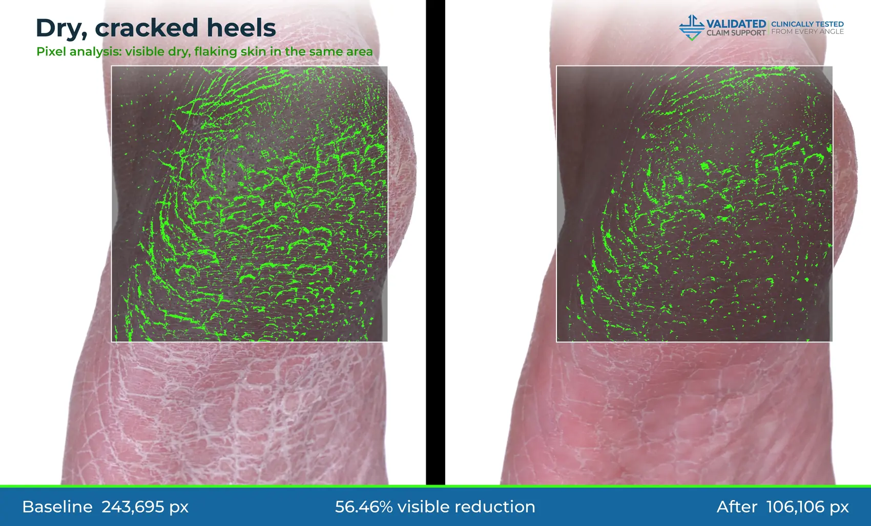
- Cracked heel: Baseline to after, 56.46% fewer flake pixels
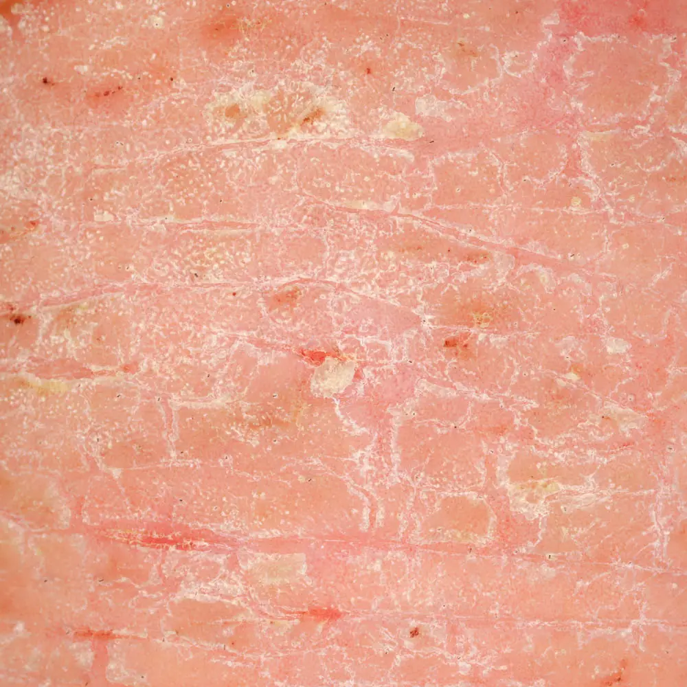
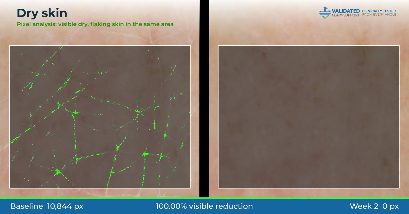
- Dry skin, close-up: Baseline to immediately after
- Eczema: Baseline to week 2
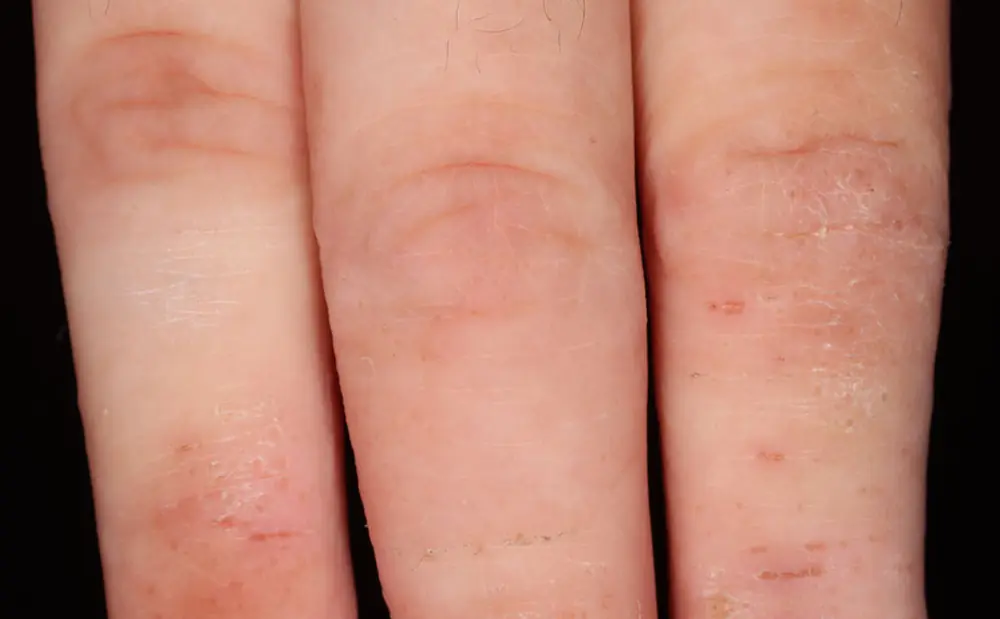
- Eczema, knuckles: Baseline to week 4
Image analysis examples
- Crow’s feet and under-eye: −37% wrinkle pixels, case study style
- Under-eye fine lines: 31.25% visible reduction, deck style
- Acne lesions: −50% lesion pixels, case study style
- Cracked heel: 72.74% visible reduction, deck style
- Redness: −79% red pixels, case study style
- Under-eye fine lines: 30.55% visible reduction, deck style
- Dry, flaking heel: −93% flake pixels, case study style
- Cracked heel: 56.46% visible reduction, deck style
- Dry skin: 100% visible reduction, deck style
Library
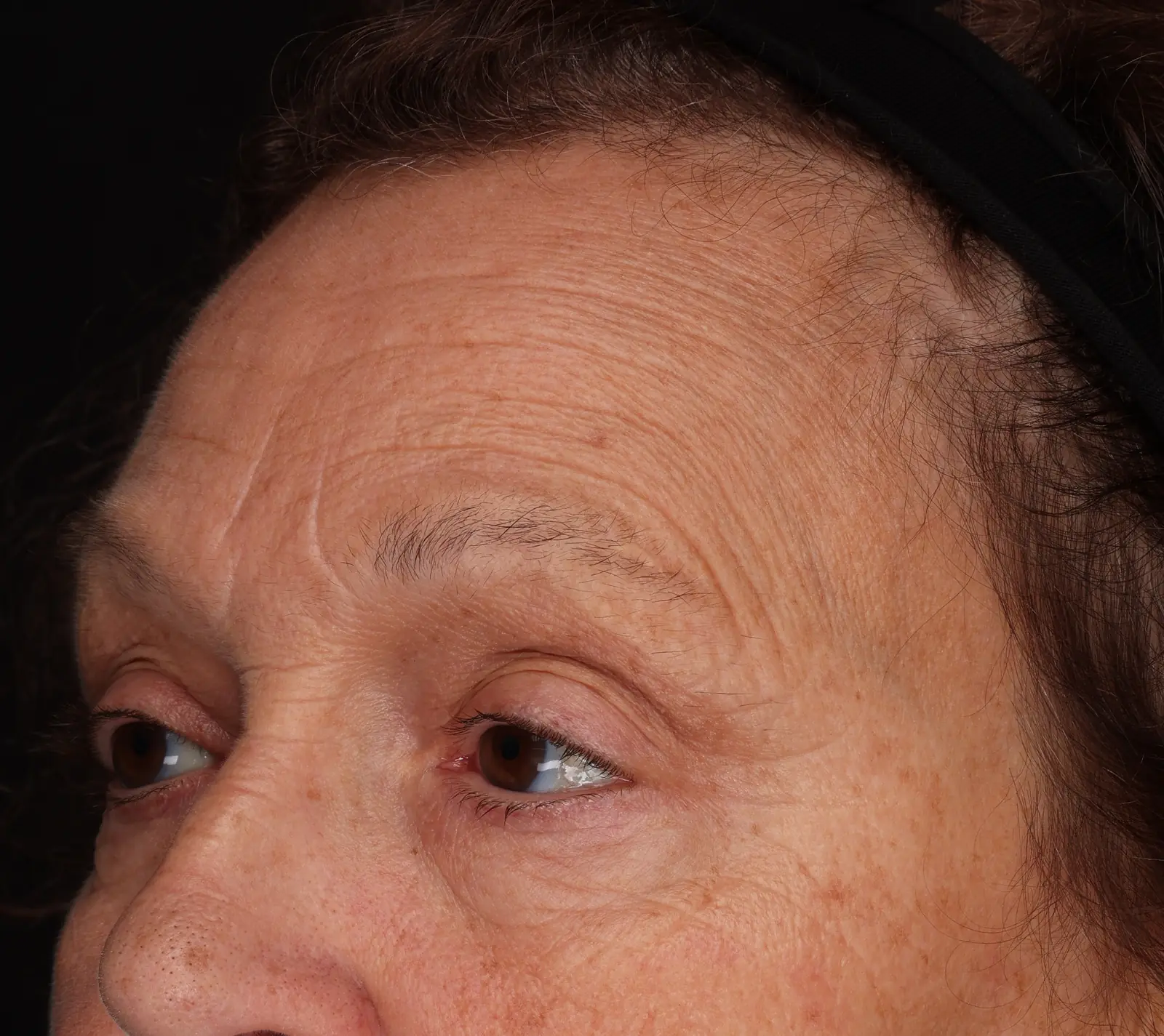
- Wrinkles, baseline to T1
- Acne, baseline to week 4
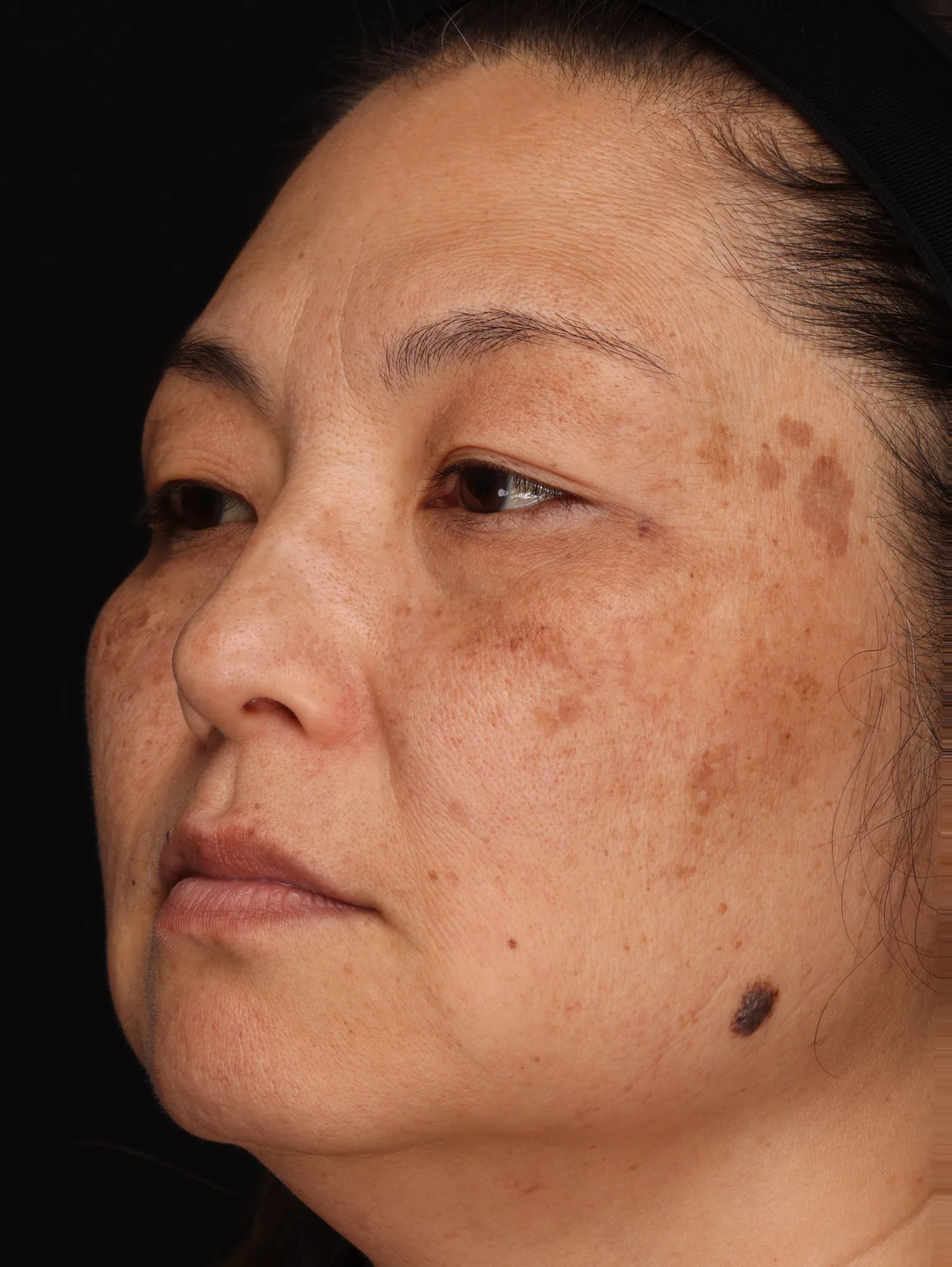
- Age spots, baseline to week 12
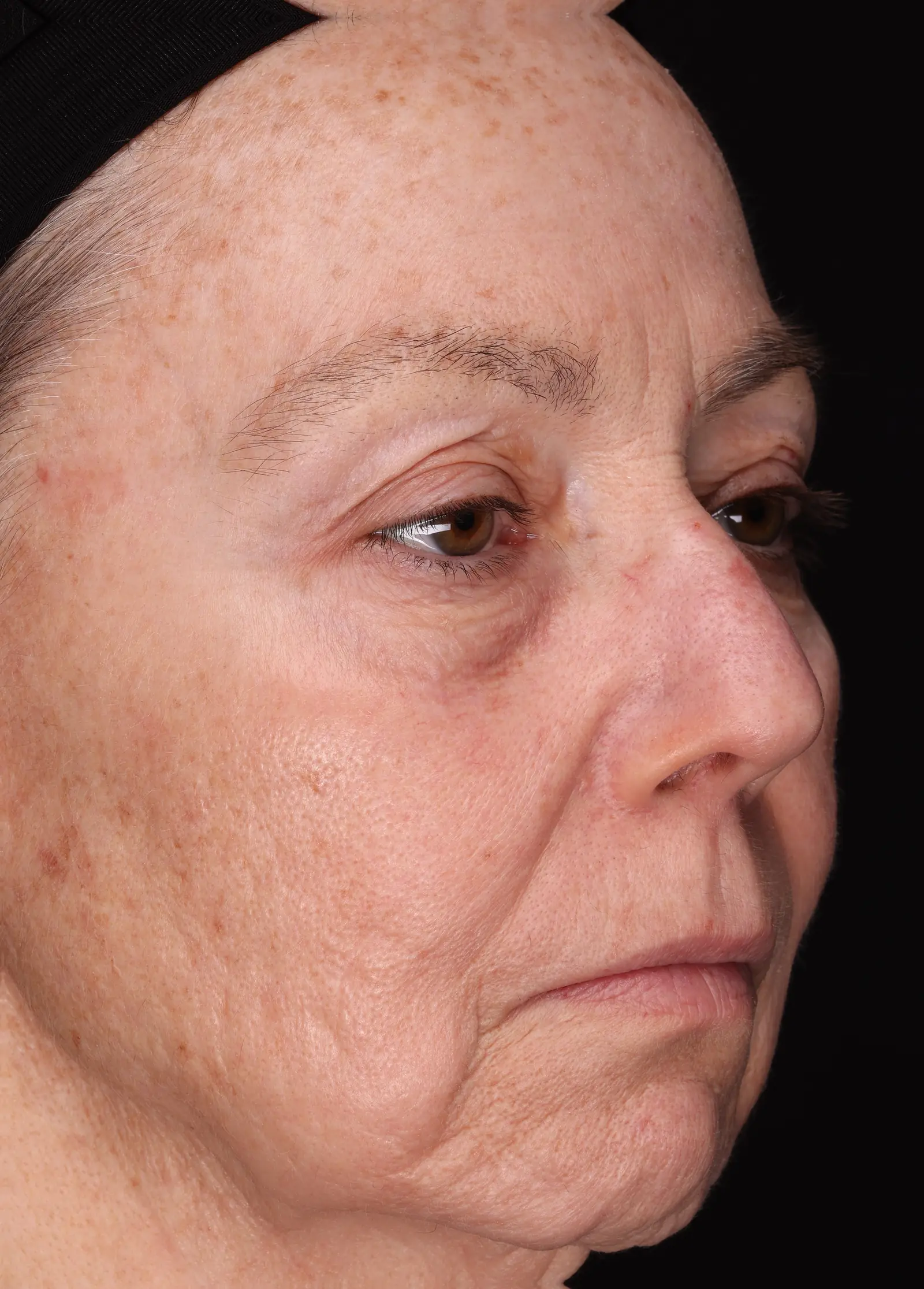
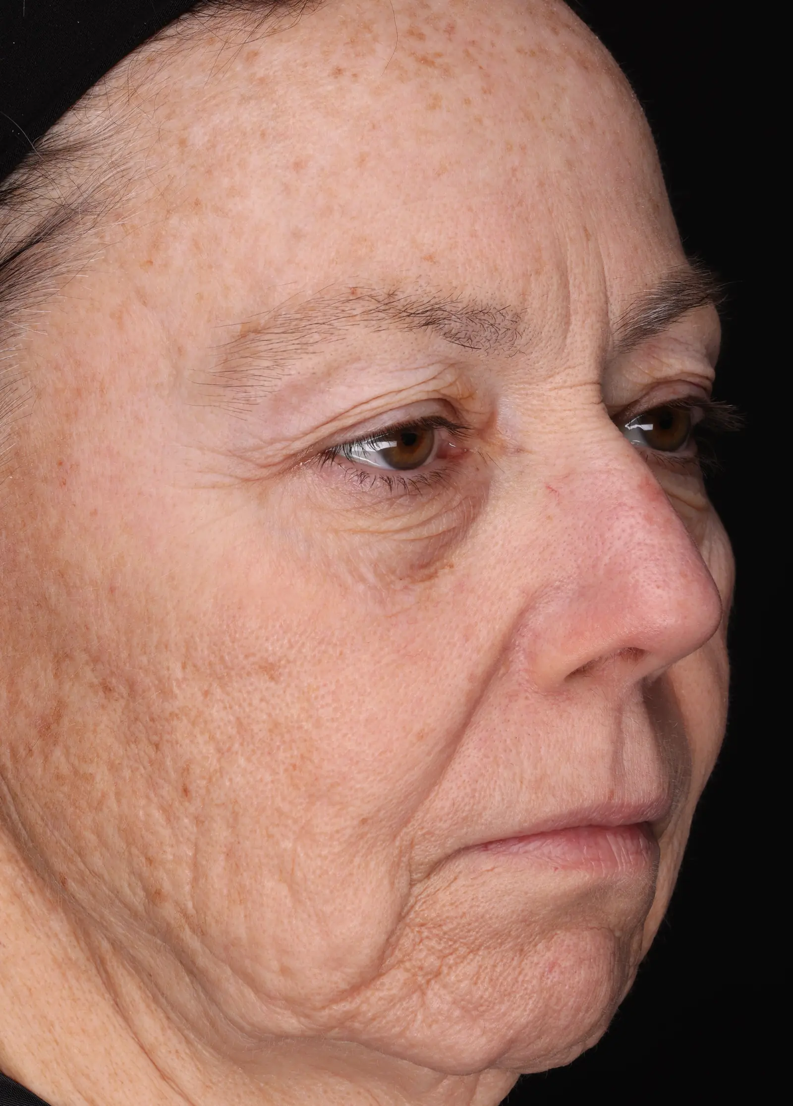
- Under-eye, nose up
- Redness, baseline to week 2
- Under-eye, baseline to week 8
- Dry sole of the foot
- Cracked heel
- Cracked heel
- Eczema, knuckles